X-ray Sources 101: What’s a Microfocus Source, Anyway?
Definition of a Microfocus Source
Much to the chagrin of many in our industry, there’s no legal (or even just widely accepted) definition of a microfocus source. This leads to some understandable confusion among the X-ray source buying population, and to some occasional overly generous prose by the marketers in some X-ray source companies.
In some X-ray market segments, a microfocus is any source with a focused X-ray spot smaller than 1mm (the thinking goes, if it has a µm in front of the spot size, it’s a microfocus). Other manufacturers feel that there’s a cutoff around 100µm in order to be considered a microfocus. Still other X-ray industry pros feel that a microfocus refers to any X-ray source that’s actively focused rather than statically focused (maybe a bit too technical to be useful). There’s no right or wrong answer, it’s not like there’s an ISO definition of microfocus that’s agreed and enforceable.
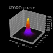
So what about Micro X-Ray? We specialize in small spots, so by some of those definitions every tube we make is a microfocus tube. But when we say microfocus we generally mean any source that focuses X-rays down to under 10µm like our Microbox integrated source, and we refer to sources with larger spot sizes as Minifocus sources. Our largest spot size sources are 1000-1500µm, and in those cases we don’t generally talk about their spot sizes at all, because in the applications they’re designed for spot size isn’t an important specification point. If you need a refresher on what exactly we mean when we’re talking about focal spot, then check out our post on the topic in the Resources section of our website.
Spot Sizes in Imaging
Imaging applications are generally all about identifying features hidden inside opaque materials. Whether you’re imaging a rock, a pebble, or a grain of sand, you can use X-rays to see inside them all. X-rays have the ability to penetrate objects that visible light cannot, which makes them incredibly valuable in a huge array of application spaces from security to food inspection to electronics manufacturing. With X-ray sources, there are three main knobs you can turn that impact the final image:
- Excitation voltage: This sets the maximum penetration ability of the X-rays. The higher the voltage, the higher density object you are able to image.
- Beam current: This sets the absolute number of X-rays from the source. The higher the beam current, the more X-rays. The more X-rays, the faster an image can be acquired, and the faster your product can be moved through the process.
- Spot size: This sets the minimum feature size able to be imaged in an X-ray system. The smaller the spot size, the smaller the size of the feature that can be resolved on the X-ray image
If we think about these three knobs, you can start to map them on to your application and narrow down your X-ray source options. If you have a general idea of the density of the object to be scanned, the process time you have available to scan it, and the size of the features you’ll need to resolve, you have a great starting point for selecting an X-ray source. Of course, there are always tradeoffs, so you might choose a slightly higher resolution that takes a slightly longer time, or perhaps the ability to have a quick process time might come at the expense of slightly lower contrast on dense materials. Either way, once you have your application properly scoped, at a great point to reach out to our technical team so we can find the right source together.
Spot Sizes in Analysis
Most analysis applications couldn’t care less about spot size. Take X-ray attenuation thickness gauging, for instance. This application works by passing a thin film of some known material between an X-ray source and an X-ray detector. The system is calibrated with the tube and detector a fixed distance apart, and the total flux from the X-ray tube is empirically measured and stored. Because the material is known, the absorption of the material for a given X-ray energy can be calculated. During operation, the X-ray flux is read back continuously from the detector, and the thickness can be calculated with a high degree of precision based solely on the attenuation of the X-ray beam through the film. At no point in this process is the spot size of the X-ray tube impactful, and for this reason the majority of thickness gauging applications tend to use tubes with our largest spot sizes, around 1000um.
Of course, not all X-ray analysis applications are created equally, and for some (I’m looking at you, µXRF) the spot size is critically important. But in general, spot size usually only matters for imaging applications.
Summary
The definition of a microfocus source matters much less than selecting the right spot for you application. If you’re in the market for an X-ray source of any kind, it’s best to understand your application and select a source with the spot size you need, and understand that terms like microfocus, small spot, etc are really all just marketing terms with no standardized definition. The spot size YOU need depends on YOUR use case. We here at Micro X-Ray are always happy to discuss your applications, and get you the right tube for your application, whether you need a 5µm spot, a 1500µm spot, or something in between.



 2020 Micro X-Ray


 2020 Micro X-Ray